Indolent Tuberous Sclerosis
Araújo EF
DOI10.21767/2577-0578.10029
Araújo EF*
Internal Medicine Residence Program Department, Centro Hospitalar São João, Portugal
- *Corresponding Author:
- Araújo EF
Internal Medicine Residence Program Department
Centro Hospitalar São João, Portugal
Tel: +351961313106
E-mail: e_filipe_araujo@hotmail.com
Received Date: January 20, 2017; Accepted Date: January 23, 2017; Published Date: January 30, 2017
Citation: Araújo EF. Indolent Tuberous Sclerosis. Insights Chest Dis. 2017, 2:2.
Abstract
Clinical Image
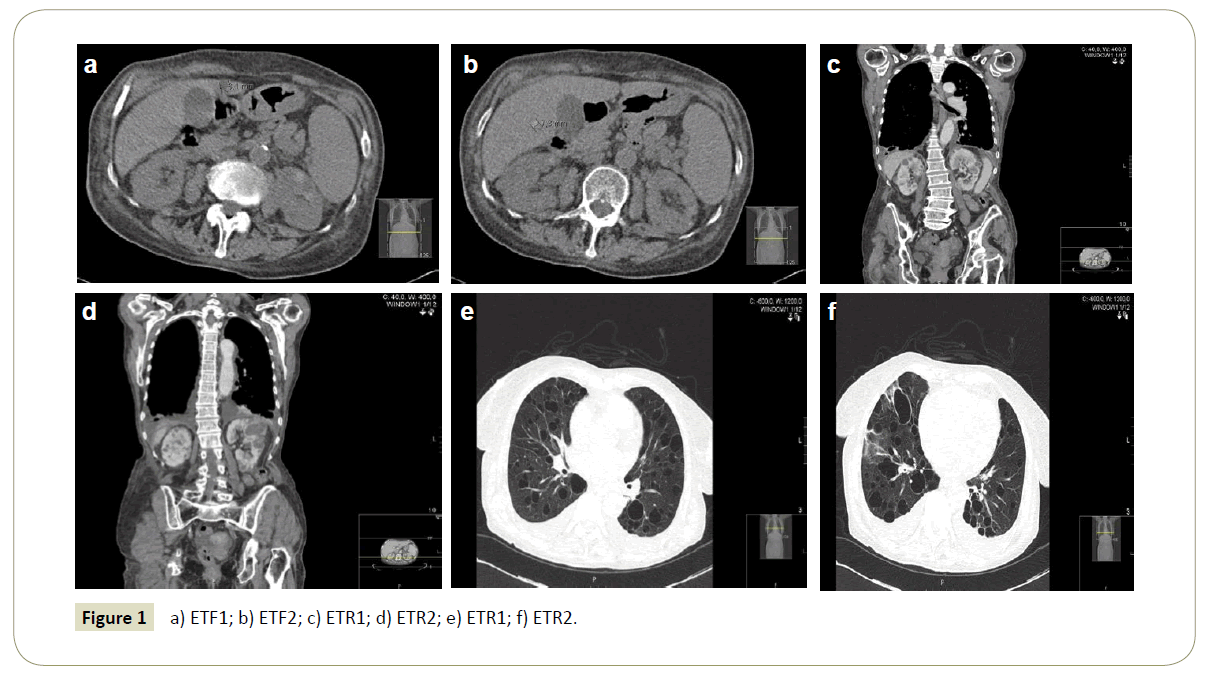
Computed Tomography (CT) images of an eighty years’ woman, with a long-standing diagnosis of asthma, admitted in hospital for Community Acquired Pneumonia with Hypoxemic Respiratory Insufficiency, without clinical improvement after antibiotic therapy and optimization of bronchodilation therapeutics. In the lung images, we can observe multiple cystic formations, of different sizes and thin wall, spread along all lung suggesting lymphangioleiomyomatosis. Abdominal CT shows hepatic cysts and multiple renal cystic formations. The association of pulmonary lymphangioleiomyomatosis and renal cysts favours the diagnosis of indolent tuberous sclerosis. The various phenotypes of the disease include forms of light disease, with normal survival rates (Figure 1).
Open Access Journals
- Aquaculture & Veterinary Science
- Chemistry & Chemical Sciences
- Clinical Sciences
- Engineering
- General Science
- Genetics & Molecular Biology
- Health Care & Nursing
- Immunology & Microbiology
- Materials Science
- Mathematics & Physics
- Medical Sciences
- Neurology & Psychiatry
- Oncology & Cancer Science
- Pharmaceutical Sciences

