Hiatal Hernia with Intra-Thoracic Communicating Hydrocele
Lim CK
DOI10.21767/2577-0578.10030
Lim CK*
Department of Internal Medicine, Division of Chest Medicine, Far Eastern Memorial Hospital, New Taipei City, Taiwan (R.O.C)
- *Corresponding Author:
- Lim CK
Department of Internal Medicine
Division of Chest Medicine, Far Eastern Memorial Hospital
No. 21, Section 2, Nanya South Road
Banciao District, New Taipei City 220, Taiwan (R.O.C).
Tel: +886-2-89667000
E-mail: ycck55@yahoo.com.tw
Received date: June 04, 2016; Accepted date: February 23, 2017; Published date: March 06, 2017
Citation: Lim CK. Hiatal Hernia with Intra-Thoracic Communicating Hydrocele. Insights Chest Dis. 2017, 2:3.
Clinical Study
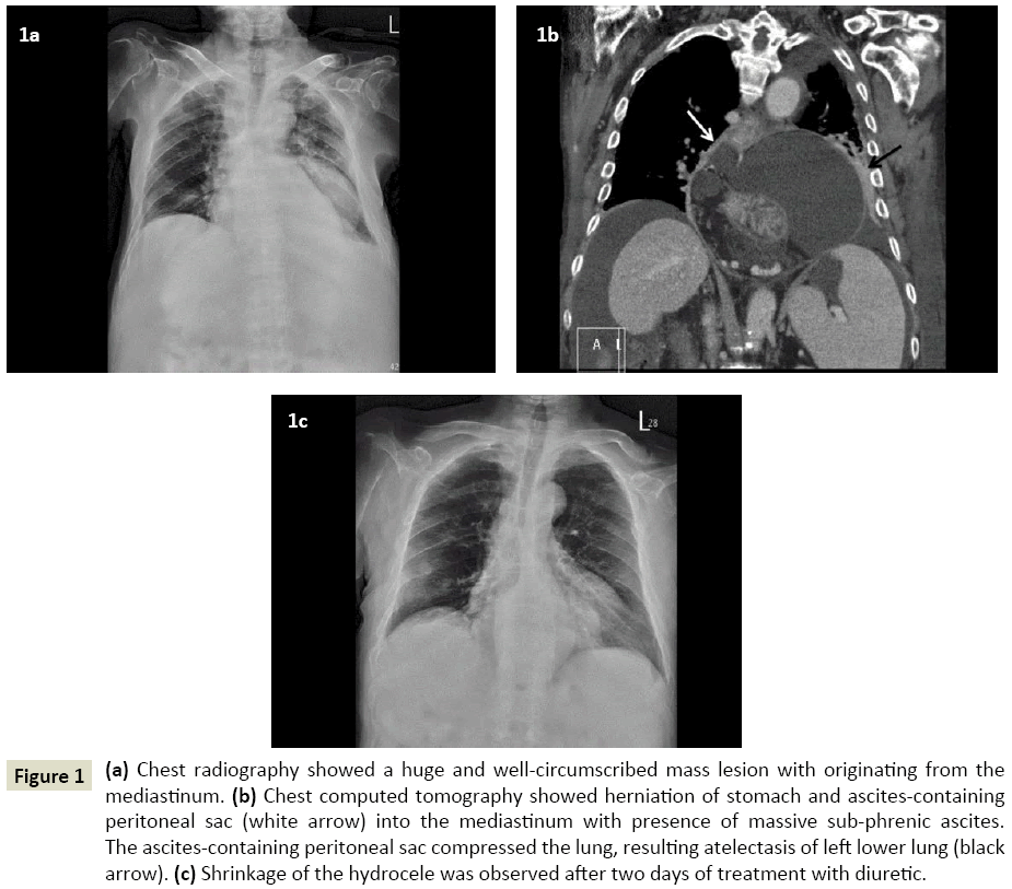
A 72-year-old man with alcoholic liver cirrhosis presented to our clinic with the chief complaints of shortness of breath and abdominal distension for one week. Upon physical examination, diffuse wheezes were auscultated bilaterally. The abdomen was distended with presence of umbilical hernia and shifting dullness. His lower limbs showed pitting edema. The chest radiography (Figure 1a) showed a huge and well-circumscribed mass lesion originating from the mediastinum. Chest computed tomography (Figure 1b) revealed herniation of stomach and ascitescontaining peritoneal sac into the mediastinum with presence of massive sub-phrenic ascites. Passive atelectasis of left lower lung was also noted, compressed by the ascites-containing peritoneal sac. Hiatal hernia with intra-thoracic communicating hydrocele was diagnosed. Furosemide and spironolactone were initiated for ascites control. Two days later, his shortness of breath and abdominal distension showed dramatic improvement. The chest radiography (Figure 1c) before discharge showed shrinkage of the hydrocele. He receives regular follow-up at hepatology outpatient thereafter for alcoholic liver cirrhosis. He also receives furosemide and spironolactone regularly for ascites control. No recurrence of the intra-thoracic communicating hydrocele since then.
Figure 1: (a) Chest radiography showed a huge and well-circumscribed mass lesion with originating from the mediastinum. (b) Chest computed tomography showed herniation of stomach and ascites-containing peritoneal sac (white arrow) into the mediastinum with presence of massive sub-phrenic ascites. The ascites-containing peritoneal sac compressed the lung, resulting atelectasis of left lower lung (black arrow). (c) Shrinkage of the hydrocele was observed after two days of treatment with diuretic.
Hiatal hernia refers to the herniation of intra-abdominal elements into the mediastinum through the oesophagus hiatus [1]. Hiatal hernia can be classified as sliding and paraesophageal, for which sliding hiatal hernia accounts for most of the cases. It is caused due the circumferential laxity or localized defect of pharyngoesophageal membrane [1].
With the presence of massive ascites, peritoneal sac may herniate into the mediastinum via gastroesophageal junction, even though there is absence of hiatal hernia [2]. This may result a mass lesion visible on chest radiography, masquerading a mediastinal mass that leads to confusion between mediastinal cysts, abscess, or pancreatic pseudocyst. Prompt recognition of this entity in patients with massive ascites is mandatory, so that unnecessary work-ups and diagnostic confusion can be avoided.
References
Open Access Journals
- Aquaculture & Veterinary Science
- Chemistry & Chemical Sciences
- Clinical Sciences
- Engineering
- General Science
- Genetics & Molecular Biology
- Health Care & Nursing
- Immunology & Microbiology
- Materials Science
- Mathematics & Physics
- Medical Sciences
- Neurology & Psychiatry
- Oncology & Cancer Science
- Pharmaceutical Sciences

