Cardiac Paraganglioma; Superiority of 18F-FDG PET/CT in Evaluating Prevalence of a Potentially Aggressive Disease in The Associated Familial Paraganglioma Syndrome
Fathinul Fikri AS, Abdul J Nordin and Zanariah H
DOI10.21767/2577-0578.10001
Fathinul Fikri AS1*, Abdul J Nordin2 Zanariah H3
1Centre for Diagnostic Nuclear Imaging, Faculty of Medicine and Health Science, Universiti Putra Malaysia 43400 Serdang, Selangor, Malaysia
2Centre for Diagnostic Nuclear Imaging, Faculty of Medicine and Health Science, Universiti Putra Malaysia 43400 Serdang, Selangor, Malaysia
3Endocrinology Unit, Department of Medicine, Hospital Putrajaya Putrajaya, 62250/ Malaysia
- *Corresponding Author:
- Fathinul Fikri AS
Centre for Diagnostic Nuclear Imaging
Faculty of Medicine and Health Science
Universiti Putra Malaysia, 43400, Serdang
Selangor, Malaysia
Tel: +03 89471646
Fax: +03 89472775
E-mail: ahmadsaadff@gmail.com
Received date: August 28, 2015 Accepted date: August 28, 2015 Published date: September 28,2015
Citation: Fathinul F AS, Nordin AJ, Zanariah H (2015) Cardiac Paraganglioma; Superiority of 18F-FDG PET/CT in Evaluating Prevalence of a Potentially Aggressive Disease in The Associated Familial Paraganglioma Syndrome. Insights Chest Dis. 1:1.
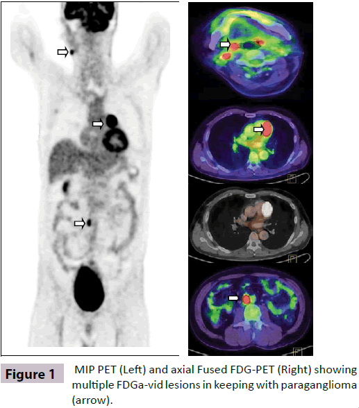
A 45-year-old male patient with an inoperable cardiac paraganglioma (PGL) and history of resected bilateral carotid paragangliomas was referred for 18F -FDG PET/CT study to exclude recurrent tumour. The patient was hypertensive with elevated urine catecholamine on a routine annual follow-up. Family history was positive for PGL [1,2]. 18F -FDG PET/CT revealed an avid 18F -FDG left supracardiac PGL with multiple new avid FDGfoci adjacent to the right internal carotid artery and in the aortocaval region denoting recurrent metachronous tumours (Figure 1). These lesions exhibited high maximum standard uptake values (SUVmax) ranging from 10 to 40. Our case highlights the importance of 18F –FDG PET/CT as a superior armamentarium in localizing recurrent PGLs in patient with mediastinal PGLs with the associated familial PGL syndrome [3,4]. They have tendency to develop metastatic disease indicating that these tumours are often aggressive and should be carefully followed.
References
- Paul S, Jain SH, Gallegos RP, Aranki SF, Bueno R (2007) Functional Paraganglioma of the Middle Mediastinum. AnnThoracSurg83:e14-16.
- Ghayee HK, Havekes B, Corssmit EPM, Eisenhofer G, Hammes SR, Ahmad Z, et al. (2009) Mediastinalparagangliomas: association with mutations in the succinate dehydrogenase genes and aggressive behavior. EndocrRelat Cancer 16:291-299.
- Turley AJ, Hunter S, Stewart MJ (2005) A cardiac paraganglioma presenting with atypical chest pain.EurJCardiothoracSurg28 :352-354.
- Timmers HJ, Kozupa A, Chen CC, Carrasquillo JA, Ling A, Eisenhofer G, et al. (2007) Superiority of fluorodeoxyglucose positron emission tomography to other functional imaging techniques in the evaluation of metastatic SDHB-associated pheochromocytoma and paraganglioma. JClinOncol25:2262-2269.
Open Access Journals
- Aquaculture & Veterinary Science
- Chemistry & Chemical Sciences
- Clinical Sciences
- Engineering
- General Science
- Genetics & Molecular Biology
- Health Care & Nursing
- Immunology & Microbiology
- Materials Science
- Mathematics & Physics
- Medical Sciences
- Neurology & Psychiatry
- Oncology & Cancer Science
- Pharmaceutical Sciences

